Study examines cardiac risk after radiotherapy for breast cancer
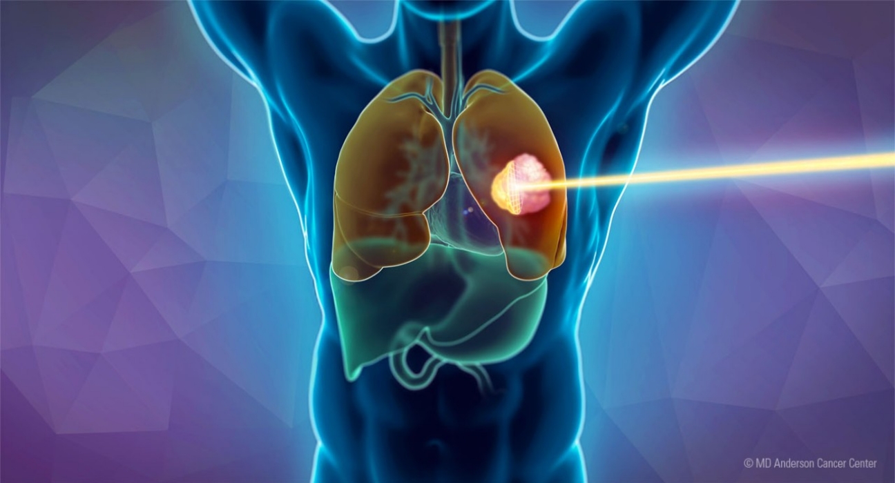
What best predicts the risk of heart damage in breast cancer patients treated with radiotherapy? A study of a Chilean cohort, with the participation of iHEALTH researchers, found that patients’ cardiovascular history carried more weight than the radiation dose received by the heart.
Radiotherapy is one of the pillars of breast cancer treatment and helps more and more women survive the disease. But this progress brings a new challenge: as survival increases, so does the likelihood of late treatment effects, including those that affect the heart.
“Post-radiotherapy cardiotoxicity encompasses the cardiac alterations that can appear after irradiating the chest, ranging from subclinical dysfunction detectable only through tests to coronary artery disease, valvular disease or heart failure,” explains Víctor Aguirre Bravo, lead author of the study, from the Radiation Safety Unit at UC CHRISTUS and the Institute of Physics at Pontificia Universidad Católica de Chile. “It is a concern because more and more women live long enough for these late effects to emerge, and heart health becomes a central part of survivorship care.”
Although treatment targets the breast, the heart lies very close to the irradiated area, especially in left-sided tumors. “It inevitably receives a fraction of the dose,” the researcher notes. “The effects are typically late: they can appear years or even decades later.”
The study, conducted by researchers from Pontificia Universidad Católica de Chile and Universitat de València, with the participation of iHEALTH Alternate Director Marcelo Andia, analyzed 42 breast cancer patients treated with radiotherapy at UC CHRISTUS, with a median follow-up of nearly seven years. The team examined the dose received by seven structures of the heart and compared it with each patient’s clinical profile before treatment.
According to the authors, this is the first Latin American cohort to link dosimetry of multiple cardiac substructures with clinical outcomes. “Most of the evidence on cardiotoxicity comes from cohorts in the Global North, and most risk models were developed in those settings. This raises questions about their applicability to populations with different epidemiological, genetic and socioeconomic realities,” says Aguirre. “Generating local evidence is essential to inform regional guidelines.”
The issue is especially relevant for Chile, where cancer is projected to overtake cardiovascular disease as the leading cause of death within the next decade.
Artificial intelligence to search for patterns
The team applied machine learning in two ways. “First, using unsupervised techniques, which look for natural groupings in the data without knowing the outcome, we explored whether patients spontaneously separated according to whether or not they developed cardiotoxicity,” Víctor explains. “Second, using supervised models, which learn from cases with known outcomes, we compared three approaches: one based on clinical variables, another on cardiac dose variables, and a third combining both.”
Ten of the 42 patients (23.8%) developed cardiovascular events. There were no differences in the dose received between those who experienced cardiac events and those who did not, in any of the seven heart structures analyzed. Pre-existing cardiovascular disease, by contrast, was the strongest predictor: it was present in 60% of patients with cardiotoxicity, compared with 18.8% of the rest. In addition, the four most serious events occurred in women with that history.
“In practical terms, baseline cardiac vulnerability outweighed cardiac dose,” Aguirre Bravo summarizes.
Does the dose not matter?
The researcher cautions that the result should not be read as meaning that radiation to the heart is irrelevant. “Not finding dose differences does not prove that dose is irrelevant. With 42 patients, the study could only detect very large associations,” he clarifies. “The correct interpretation is that, with low doses and patients with a high burden of comorbidities, the risk contributed by the dose received is overshadowed by baseline cardiovascular risk.”
The team also evaluated the American Heart Association’s (AHA) PREVENT score, which estimates cardiovascular risk using routine data from the medical record, such as age, blood pressure, lipids, kidney function and diabetes. Although it did not show a statistically significant risk gradient, it was much more stable than the dose-only models, so the authors propose it as a hypothesis to be tested in larger cohorts.
“Because this is an exploratory study, we are not proposing clinical recommendations, but hypotheses,” Aguirre Bravo emphasizes. “The main one is that assessing baseline cardiovascular risk with data already in the medical record could be the most solid starting point for identifying patients at highest risk.”
His message for patients is straightforward: “Radiotherapy remains a fundamental treatment, and it is important to inform the care team of any history of heart conditions.”
Next steps include larger, prospective cohorts, validation at other centers and structured cardiology follow-up. “Another key aspect is quantifying biases in dose determination for cardiac substructures, which depend, for example, on how small structures such as the left anterior descending artery are contoured. That is precisely one of the subprojects we are developing,” the researcher anticipates. “Our contribution is a replicable protocol to move toward this type of study in the region.”
Aguirre-Bravo V, El Far R, Herrera Ruiz M, Gálvez Santelices S, Muñoz-Schuffenegger P, Merino T, Diez Domingo S, Andia ME. Machine learning predictors of post-radiotherapy cardiotoxicity in a Latin American breast cancer cohort: Clinical risk factors outperform cardiac dosimetry. Journal of Applied Clinical Medical Physics, 2026;27:e70862. DOI: 10.1002/acm2.70862
