How close are we to simulating a knee fracture on a computer?
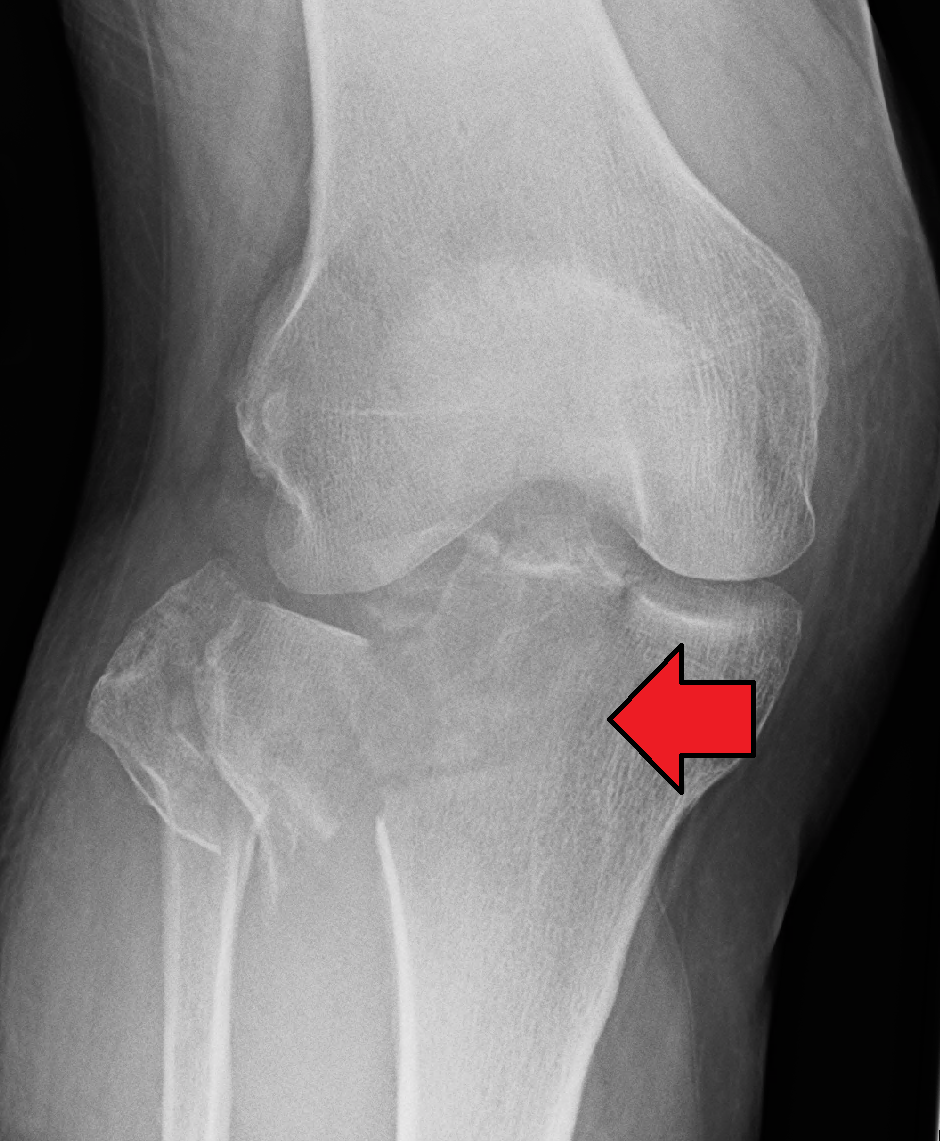
A systematic review analyzed 31 studies that use computational models to evaluate tibial plateau fracture surgery. The authors conclude that the tool holds great potential, but still has a way to go before it reaches the operating room.
Tibial plateau fractures affect the upper part of the tibia, right where the knee joint forms. They account for 1.2% of fractures in adults and are the most common bone injury of the knee. They occur in older people after low-energy falls and in younger people after high-impact accidents. In the latter cases, they often require surgery, in which the bone fragments are fixed with plates and screws. Although techniques have improved, many patients are left with lasting effects: reduced mobility, sick leave lasting more than a year, and an overall perception of poor outcomes.
To improve these treatments, biomechanical research needs to test how different implants perform. Ideally, this is done on human cadaver bones, but that is expensive, each specimen can only be used for a single test, and it involves complex ethical and safety requirements. That is why finite element modeling, an engineering technique that allows these tests to be carried out on a computer, is increasingly being used.
A team from Pontificia Universidad Católica de Chile, Fundación Epistemonikos, Universidad del Desarrollo, Universidad de los Andes, Universidad Técnica Federico Santa María and iHEALTH systematically reviewed everything published on the use of this technique in tibial plateau fractures. The study was published in the Journal of the Mechanical Behavior of Biomedical Materials.
“Finite elements are an example of interdisciplinary work that caught our group’s interest, and to launch this line of research, we felt it was important to have a clear picture of the state of the art in applying this method to a relevant condition within our subspecialty,” explains orthopedic surgeon Pablo Besa, first author of the study.
Breaking a big problem into many small ones
What is a finite element model? “It lets you break down complex mechanical problems into smaller ones and solve them one at a time. The sum of all those smaller results then solves the system as a whole,” Besa summarizes.
In practice, the bone, the fracture and the implants are reconstructed in three dimensions, often from patients’ CT scans. The model is then divided into thousands of tiny pieces, and the response of each one is calculated when the knee bears a load—for example, while standing. This makes it possible to control and observe every variable, repeat the test as many times as needed at low cost and, in theory, create simulations tailored to each patient.
The team reviewed more than 700 articles and selected 31 studies published between 2011 and 2025. The results show a growing field, but one with significant limitations. Most studies created the fracture on the computer rather than using real patient cases, and almost none simulated the dynamic loads produced during walking. Only one compared its results with real mechanical tests. In addition, each study used different assumptions to represent the bone and the implants, making it difficult to compare them with one another.
To assess study quality, the authors used ROBFEAD, a tool originally developed to evaluate this type of model in dentistry. Under that criterion, all 31 studies were rated as having a high risk of bias. Regarding its use in orthopedics, Besa explains that “rather than adaptations,” what is needed is “to validate that its use in orthopedics has good inter- and intra-observer agreement”—that is, that different evaluators, or the same evaluator at different times, reach the same conclusions. It must also be confirmed “that it can distinguish the methodological and reporting quality of the studies.”
How far are these simulations from what actually happens in a real patient’s knee? “Quite far, but that doesn’t mean the results are useless or any less valid,” Besa clarifies. “What matters is the question you ask the models, and then determining whether the model’s fidelity is preventing you from answering that question.”
He gives an example: “If my goal is to compare two fixation techniques, the simplification may have little effect on the comparison. What that low fidelity affects is the ability to ask more complex, patient-specific questions.”
That is precisely the long-term goal: for a surgeon to be able to simulate a specific patient’s fracture before operating. How far off are we? “Very far, but we’re heading there,” he replies. “We still lack basic science knowledge to bring the models closer to what’s happening in real life. That step is crucial, and the work we’ve done brings us closer to understanding the limitations and filling the knowledge gaps one at a time.”
Besa P, Vidal C, Ramos-Rojas JT, Goldbaum E, Andia ME, Mura J. Finite element analysis in tibial plateau fractures: a systematic review of literature. Journal of the Mechanical Behavior of Biomedical Materials 182 (2026) 107530. https://doi.org/10.1016/j.jmbbm.2026.107530
