Cardiac MRI in a single breath-hold: iHEALTH researchers image the entire ventricle in about 15 seconds
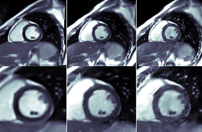
Published in Magnetic Resonance in Medicine, the study combines a low-field (0.55 T) MRI scanner with a self-supervised neural network, called ST-DIP, that jointly reconstructs eight slices of the heart acquired while the patient holds their breath just once.
Cardiac magnetic resonance imaging is the gold standard for assessing heart function. One of its measurements, known as cine, shows the heart in motion and makes it possible to calculate key indicators such as left ventricular ejection fraction. However, obtaining these images requires considerable effort from the patient.
“Typically, between 8 and 12 slices are acquired, and the patient holds their breath for about 15 seconds per slice, with rest periods in between. This part of the exam can take several minutes,” explains Rafael de la Sotta, research engineer and first author of the paper. Repeated breath-holds affect both the patient and the diagnostic process. “Holding your breath several times can be uncomfortable, especially for people with respiratory problems. In addition, small changes in position between breath-holds can misalign the slices and affect the measurements,” he notes.
The team worked with a 0.55-tesla scanner, a lower field strength than the 1.5 T or 3 T systems more commonly used in clinical practice. “Low-field scanners are usually more affordable, offer more space for the patient, and produce fewer distortions caused by metal implants. This can improve access and comfort,” says De la Sotta. The trade-off is technical. “The main challenge is that they capture a weaker signal, which makes it harder to obtain sharp images. That’s why we developed advanced reconstruction methods,” he adds.
The new method reduces the acquisition time for each slice to just 1.5 seconds, making it possible to capture eight slices—from the base to the apex of the left ventricle—in a single scan lasting 12 to 15 seconds. “We then reconstruct the slices jointly, obtaining images of the beating heart with quality comparable to much longer acquisitions,” Rafael summarizes.
Artificial intelligence is the key. “A trained model reconstructs the images from the scanner data. The network is self-supervised because it learns using data from the exam itself, without needing a large external database. This is very useful, because gathering thousands of MRI scans to train a network can be difficult and costly,” he explains.
ST-DIP (slice- and time-dependent deep image prior) is an extension of an earlier method, TD-DIP. “TD-DIP reconstructed each slice separately. With ST-DIP, we use a single network to reconstruct all eight slices jointly, taking advantage of the information they share. This reduced reconstruction time from about 45 to 16 minutes,” De la Sotta details.
Results comparable to an exam ten times longer
The method was evaluated in 10 healthy volunteers and compared with TD-DIP and with conventional reconstruction techniques (GRASP and SENSE). The left ventricular ejection fraction obtained with ST-DIP (58.7%) was comparable to that of a reference exam acquired at 15 seconds per slice (59.1%), with a total duration of more than three minutes. In addition, ST-DIP achieved significantly higher edge sharpness than GRASP and slightly higher than TD-DIP.
The authors caution that the sample is small and consists mainly of young people, so broader validation is needed. “The next step is to test the method in more volunteers and in patients to assess its clinical usefulness. We can also keep improving it to reduce acquisition and reconstruction times, and incorporate automatic analysis of cardiac motion,” De la Sotta concludes.
This research was carried out in collaboration with Pontificia Universidad Católica de Chile PhD students Tabita Catalán and Carlota Rivera, together with academics Francisco Sahli Costabal, René Botnar, and Claudia Prieto, principal investigators at iHEALTH.
R. de la Sotta, T. Catalán, C. Rivera, F. Sahli Costabal, R. M. Botnar, and C. Prieto, “Whole-Ventricle Cardiac Cine MRI in a Single Breath-Hold With Slice- and Time-Dependent Deep Image Prior at 0.55T,” Magnetic Resonance in Medicine (2026): 1–13, https://doi.org/10.1002/mrm.70635.
