Artificial intelligence to measure blood flow
In his thesis for the degree of Doctor in Engineering Sciences at the Pontificia Universidad Católica de Chile, Javier Bisbal developed three deep learning methods aimed at making the analysis of 4D flow magnetic resonance imaging more reliable, faster and more automated. This technique has great potential for assessing cardiovascular and cerebrovascular diseases.
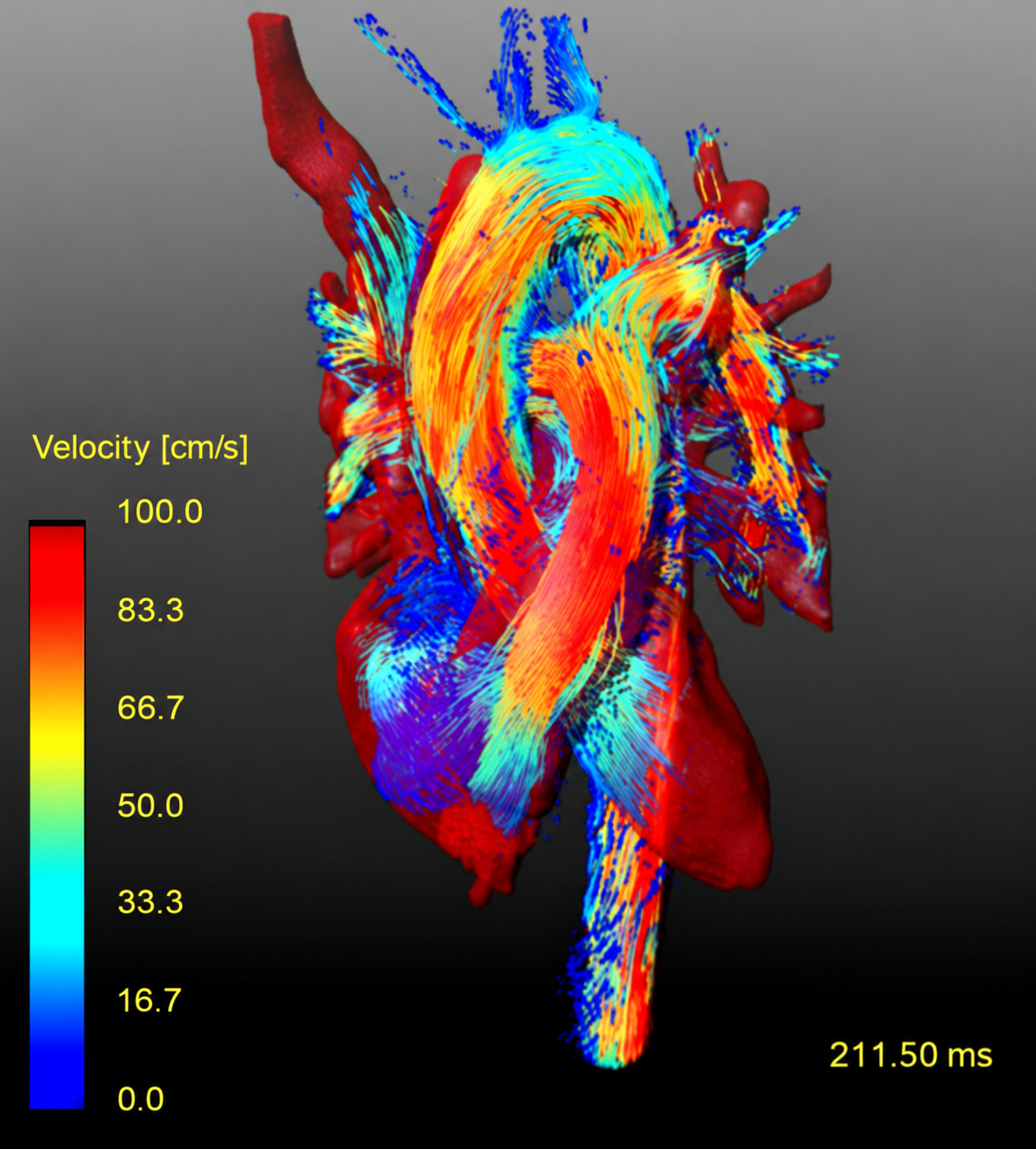
“4D flow MRI is a special magnetic resonance technique that, instead of visualizing what is static in our body, can see what is in motion. In the human body, this allows us to see how blood moves and study how it is distributed throughout the body,” Bisbal explains.
The technique is especially useful in congenital heart disease, such as aortic coarctation, in which a section of the aorta narrows. “4D flow MRI makes it possible to accurately quantify the degree of blood flow alteration and, with that, guide treatment as well as post-surgical follow-up,” he notes. In the brain, it can help characterize the degree of arterial stenosis and how blood flow is redistributed, which helps predict the risk of stroke.
Despite its potential, the technique is rarely used in clinical practice. On the one hand, acquisition is slow, since it requires collecting a large amount of three-dimensional data. On the other, obtaining diagnostically useful values requires multiple processing steps, such as noise reduction, artery segmentation and plane reformatting. “All these steps require extensive experience with image analysis software and in-depth knowledge of both the technique and the anatomy of different pathologies,” the researcher says. He adds that, according to a recent study, these limitations make the technique economically unviable outside large healthcare centers that are also engaged in research.
On top of this are noise and errors in the measured velocities. These may result from acceleration techniques, patient motion, or blood velocity exceeding the set range. In the latter case, velocity may be represented incorrectly, even appearing to flow in the opposite direction.
Improvement through deep learning
On October 9, Javier Bisbal Zenteno defended his thesis for the degree of Doctor in Engineering Sciences at the Pontificia Universidad Católica de Chile, at the San Joaquín Campus. The thesis was titled “Enhancing Hemodynamic Quantification in 4D Flow MRI Using Deep Learning.”
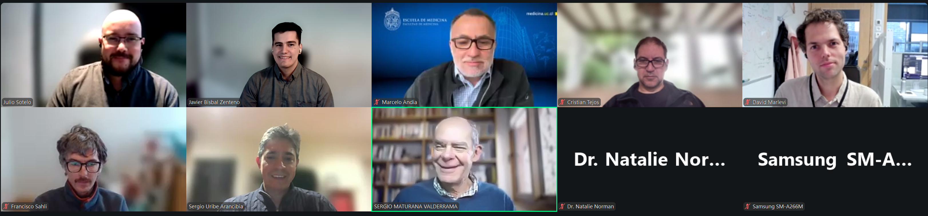
The work was supervised by professors Cristián Tejos (UC) and Sergio Uribe (Monash University). The committee also included Francisco Sahli (UC), Marcelo Andia (UC), Julio Sotelo (UTFSM) and David Marlevi (Karolinska Institutet), and was chaired by Sergio Maturana (UC), on behalf of the program director.
“I chose this topic because there was a great opportunity to bring together advances in artificial intelligence to overcome the limitations of 4D Flow and bring it closer to clinical use,” he says.
To address these challenges, the thesis develops three methods. The first corrects the measured velocities: it incorporates physical principles to reduce noise and correct errors. The second automatically identifies the planes used to quantify flow. The third labels the cerebral arteries and flags the regions where its predictions are less reliable. The methods were evaluated using simulations and data from patients and volunteers. Their results were compared with manual measurements and processing, which in some cases were performed by more than one operator to measure inter-operator variability.
According to Bisbal, automating plane reformatting could save between 20 and 30 minutes per patient, and automatic artery labeling a similar or greater amount of time in complex cases. “I had the opportunity to watch a doctor analyze a 4D flow MRI of a patient with congenital heart disease at Hospital Barros Luco Trudeau. He told me that, on average, it took him about 30 to 40 minutes to do the analysis with the planes,” he recalls. Both methods also aim to reduce variability, with predictions that take less than a minute in total.
“The idea is to support radiologists with these tools, so that AI assists them with the processing, they review it, and can easily extract metrics,” he explains. For patients, the goal is for more people to have access to this technique and to receive more complete and personalized radiology reports that help better characterize cardiovascular diseases and prevent deaths or complications.
To reach the clinic, the methods, validated in cohorts of dozens of patients and volunteers, will need to be tested in groups of hundreds of people with multiple pathologies and obtain approval from regulatory agencies. The next step is to incorporate these tools into image analysis software and carry out pilot validations in new cohorts. Along these lines, Bisbal will collaborate on the AI component of a FONDEF project awarded to Dr. Julio Sotelo to develop in-house analysis software.
The tools can also be extended to other domains. Together with Sofía Farfán, an iHEALTH PhD student, Bisbal adapted the reformatting method to low-field cardiac MRI, work they recently submitted to a conference.
In the coming weeks, Bisbal will travel to Sweden to begin a postdoctoral position at the Karolinska Institutet, in a department located within Karolinska University Hospital. “I will collaborate directly with medical staff, with the aim of bringing AI methods closer to the clinical setting,” he anticipates.