Seminar September 5TH, 2023
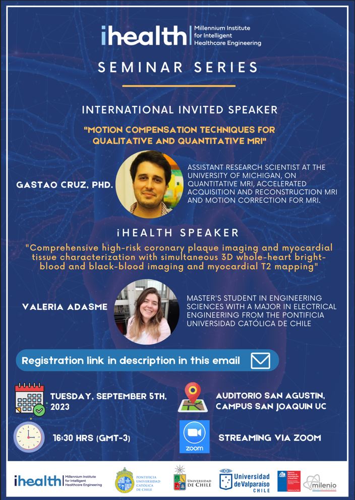
Hours: 16:30 - 18:00
Valeria Adasme
Title: Comprehensive high-risk coronary plaque imaging and myocardial tissue characterization with simultaneous 3D whole-heart bright-blood and black-blood imaging and myocardial T2 mapping.
Abstract: A combined method for Cardiac Magnetic Resonance Angiography and vessel wall imaging was proposed by Milotta, et al (2019) for assessment of coronary lumen stenosis and coronary high-risk plaque. However, this method doesn’t provide information on the degree of myocardial ischemia, edema, and necrosis. An extension using quantitative T2 map is proposed in this study to characterize the myocardium and monitor the progression of patients with Coronary Artery Disease (CAD). Using a dictionary generated with Extended Phase Graphs (EPGs) with fixed T1, we can have a good estimation of the T2 map only using the two contrasts obtained from iT2prep-BOOST. Accuracy and fixed T1 dependency were tested with a standardized T1/T2 myocardial phantom and diagnosis assessment was tested in 15 patients with suspected NSTEMI. Phantom T2 map presented an error range of ±3ms for T1 near to physiological myocardial values and low linear correlation (R2=0.97) with the fixed T1, with a low bias of -3.3ms. Patients T2 maps presented a comparable visual appearance with the anatomic data and the patients with infarcted tissue presented a T2 comparable with previously reported (T2myo=60±7ms vs T2infarcted=58.5±5.8ms (O’Brien et al., 2022)). Differentiation between infarct, myocarditis, edema, and healthy tissue was statistically significant (pvalue<0.001), except for acute infarct and edema, whose diagnosis should be determined in conjunction with the bright-blood database. Further studies are now needed to include healthy volunteers and compare with other common diseases in patients with CAD. Overall, proposed method accomplished good T2 quantification in phantom and in vivo experiments, enabling free-breathing 3D whole-heart T2 mapping with high isotropic resolution (1.2mm), predictable scan time (≈10min) and without the need of using ionizing radiation or contrast agents. This offers a tool for risk stratification, guidance or monitoring of treatment in patients with CAD and it may have great potential for clinical application.
Bio: Valeria is an electric civil engineering student at Pontificia Universidad Católica de Chile, with a major in biomedical engineering and a minor in biomedical specialization. She has a master in engineering sciences in the field of electrical engineering also at Pontificia Universidad Católica de Chile.
Gastao Cruz
Title: Motion compensation techniques for qualitative and quantitative MRI
Abstract: Patient motion is one of the primary sources of artefacts in MRI, requiring dedicated acquisition and reconstruction strategies. Respiratory, cardiac and bulk motion can all significantly impact the diagnostic quality of the scans, have different challenges and different solutions. For respiratory motion, for example, the simplest solution is asking the patient to hold their breath; however, this limits scan time (to less than twenty seconds) and not all patients will be able to comply. An alternative approach is to acquire the scan free-breathing, but monitor the breathing pattern (with a so-called navigator) and accept only data in the end-expiration phase (known as respiratory gating). Unfortunately, approximately half the data will be rejected (needing to be re-acquired), which will double the scan time. A third solution would be to acquire data free-breathing, and then incorporate the respiratory motion into the image reconstruction problem, bypassing the limitations of breath-hold and gating. In this talk, we'll look at different strategies to manage motion in MRI and how they have been applied to both qualitative and quantitative MRI.
Bio: Gastao did his PhD at King's College London (2016), working with Prof. David Atkinson, Prof. Tobias Schaeffter and Prof. Claudia Prieto in novel methods for motion corrected reconstructions in qualitative abdominal and cardiac MRI. From 2016 to 2022, he continued his work at King's as a post-doctoral researcher, now with Prof. Rene Botnar and Prof. Claudia Prieto, now focusing on motion correction methods in quantitative MRI. Currently he his an Assistant Research Scientist at the University of Michigan, where his research focuses on quantitative MRI, accelerated acquisition and reconstruction MRI and motion correction for MRI.