Seminar March 28th, 2023
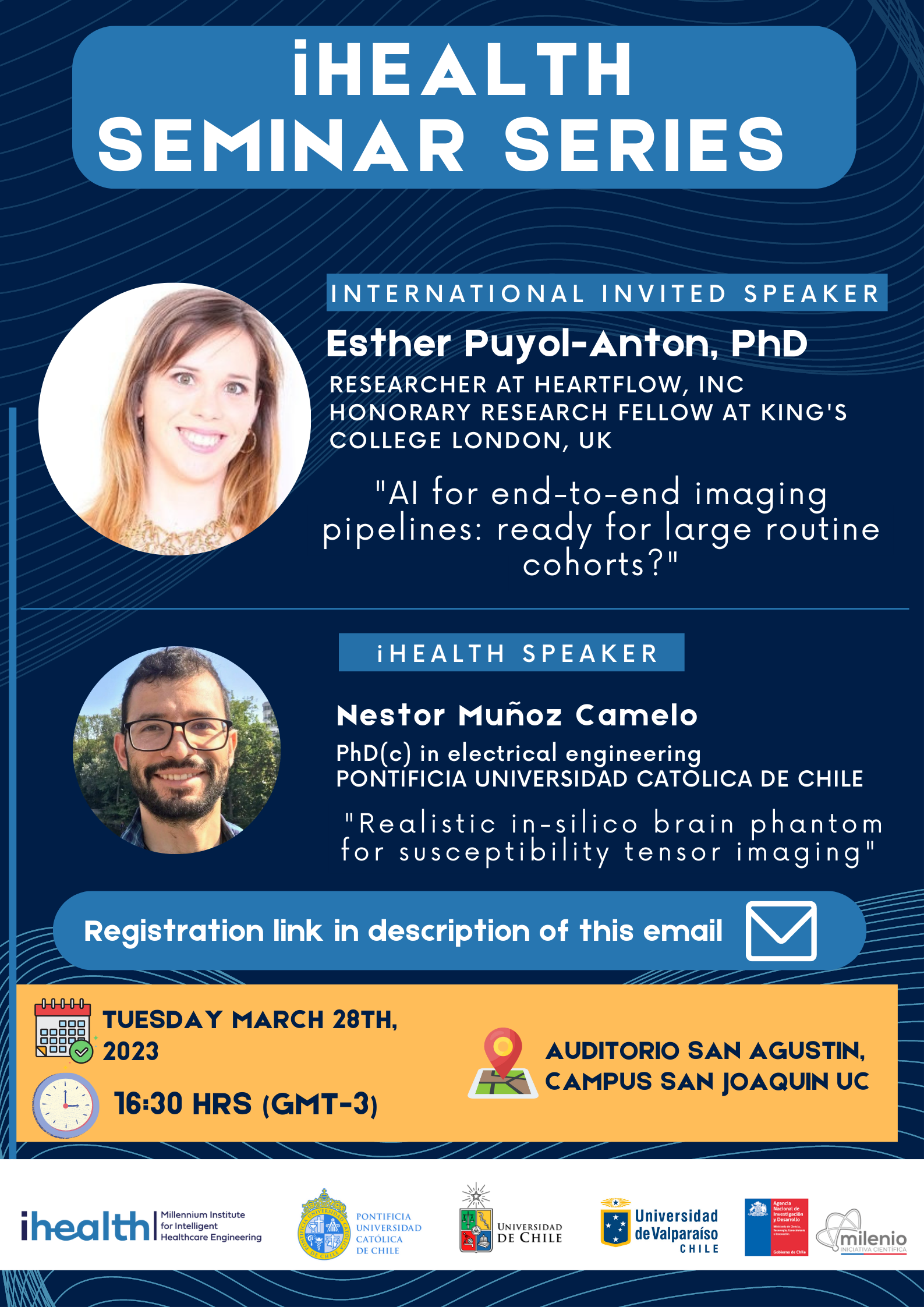
Hours: 16:30 - 18:00
Esther Puyol Anton
Title: AI for end-to-end imaging pipelines: ready for large routine cohorts?
Abstract: Cardiac magnetic resonance (CMR) plays a central role in diagnosis and management decisions for patients with heart failure (HF). However, most currently used CMR biomarkers only paint a coarse picture of cardiac health, ignoring important factors such as regional wall motion, shape, loading conditions and vascular health. A richer and more comprehensive set of biomarkers of cardiac function can be estimated from CMR but extracting them manually is unrealistically time-consuming.
The aim of our project is to improve doctors’ ability to assess heart function from CMR imaging using artificial intelligence. This talk will focus on how to use quality control and generalizable, interpretable AI models to train robust tools to automatically collect a large set of new biomarkers from cine CMR images to inform diagnosis and treatment decisions by clinicians. Final part of the talk will focus on the topic of fairness in AI and how this can affect clinical translation and the different bias strategies that can be applied.
Bio: Dr Esther Puyol Anton completed her Bachelors and Master of Science at the Polytechnic University of Catalonia (Spain) in Biomedical Signal Processing in 2014. During her studies in Spain, she enrolled in a double degree programme with Telecom Bretagne (France), where she obtained the French Master of Engineering and a Research Master in medical Imaging in 2014. Between 2014-2018, she was a PhD student in the Biomedical Engineering department at King’s College London under the supervision of Dr Andrew King, Dr Paul Aljabar and Prof Julia Schnabel from KCL, and Dr Paolo Piro from Philips Healthcare. The main aim of her PhD was to develop a multimodal statistical atlas of heart function from cardiac MR and ultrasound imaging, which could be applied using only low-cost ultrasound imaging. During the last 4 years, she was a Post-doctoral research fellow at the School of Biomedical Engineering and Imaging Sciences at King’s College London. Her main focus is developing new interpretable and quality-controlled AI systems for clinical application. Currently, she works at HeartFlow as a senior research scientist.
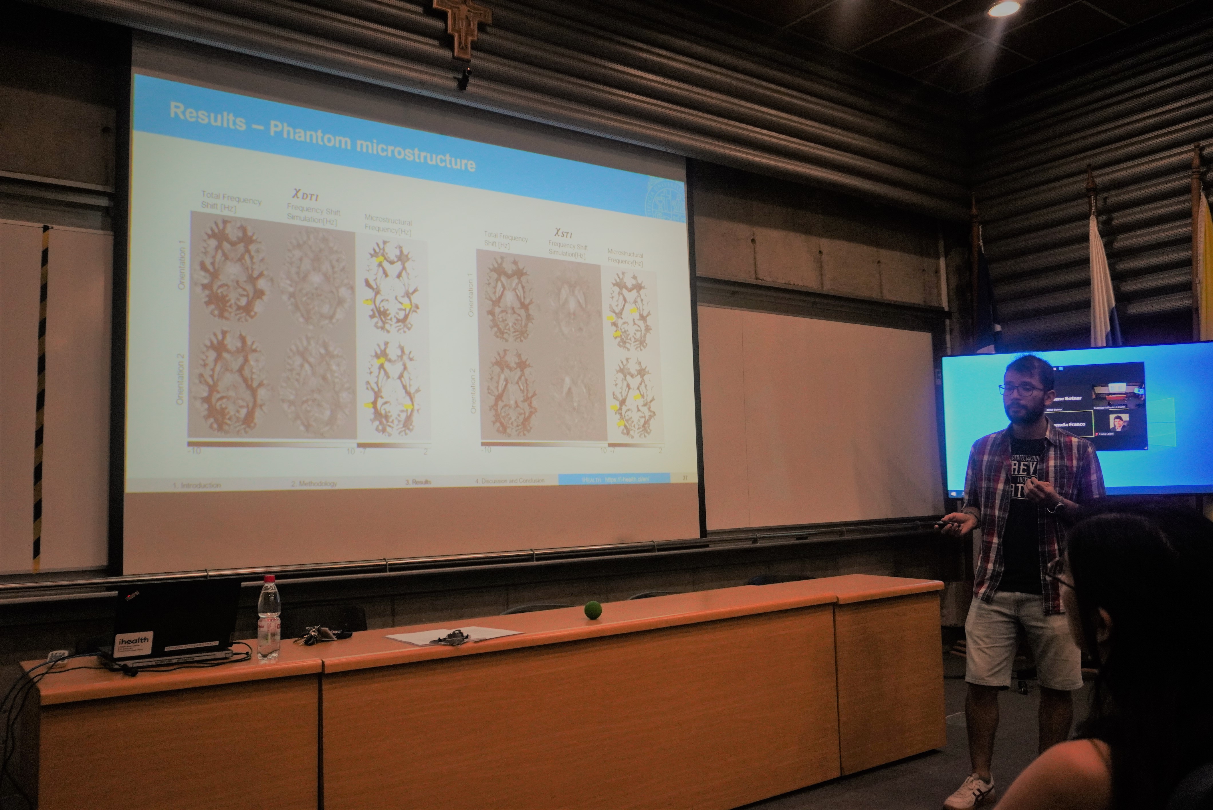
Nestor Muñoz Camelo
Title: Realistic in-silico brain phantom for susceptibility tensor imaging
Abstract: Magnetic susceptibility is the response of a material to a magnetic field. In the case of biological tissue, the magnetic susceptibility depends on the orientation of the scanned tissue, due to complex materials such as myelin in the brain. Susceptibility Tensor Imaging (STI) is a recent technique that is able to calculate the magnetic susceptibility anisotropy in the scanned tissue. Recent techniques include direct methods, such as Least Squares, or iterative methods by assuming anisotropy only in white matter. However, scanned time or computing time might be too expensive with existing algorithms. In addition, each algorithm produces different results, so it is very difficult to evaluate the performance of each algorithm. In this presentation, the design of a realistic susceptibility tensor brain phantom will be shown, including its magnetic susceptibility anisotropy behavior. This susceptibility tensor brain would work as ground truth for new and existing algorithms of STI reconstruction.
Bio: Nestor studied electronic and biomedical engineering at Universidad de Los Andes, Colombia. He is a Ph.D. candidate at Pontificia Universidad Católica de Chile, and he is currently working on the reconstruction of the Susceptibility Tensor Imaging in the brain using Deep Neural Network algorithms.