Seminar April 25th, 2023
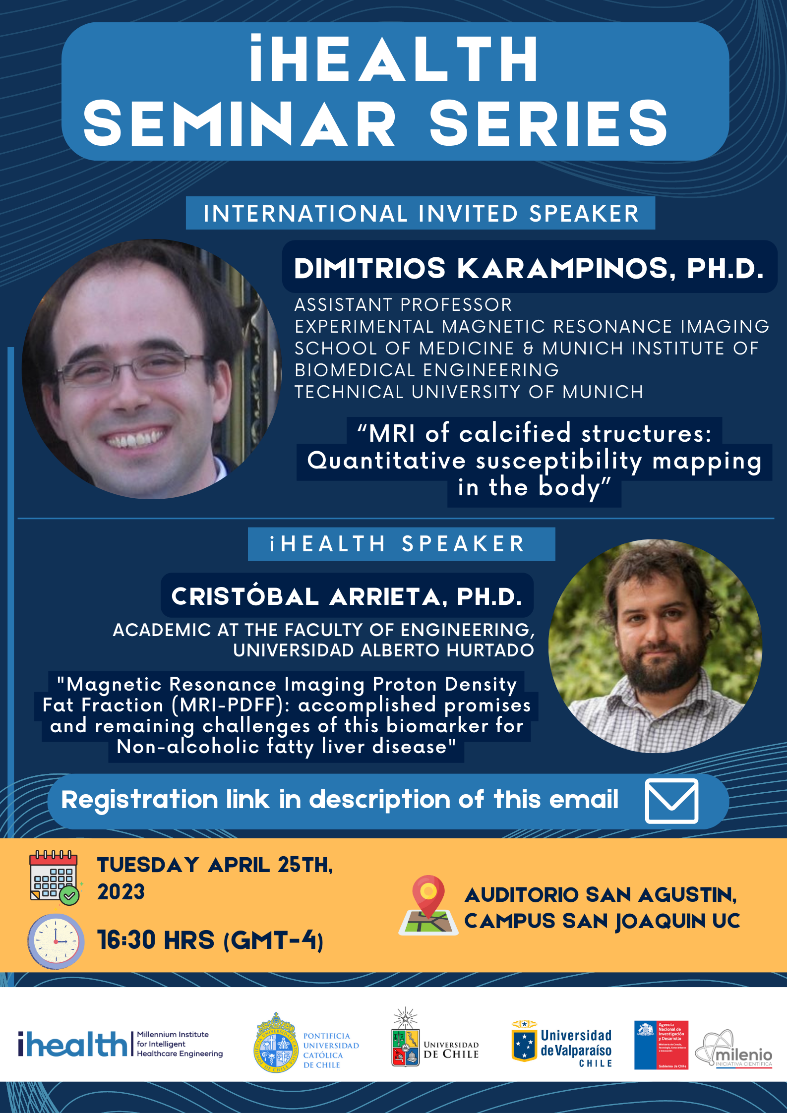
Hours: 16:30 - 18:00
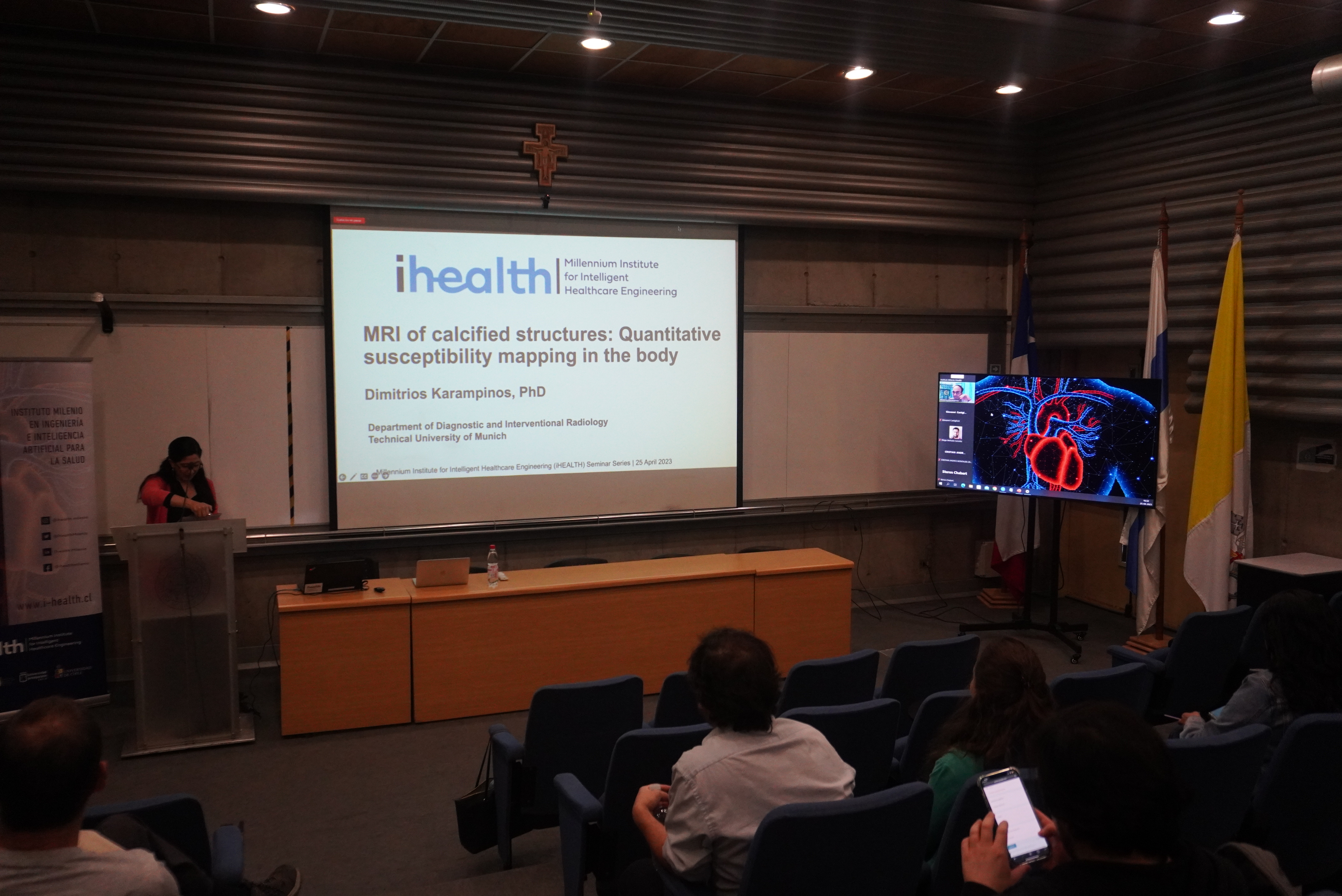
Dimitrios Karampinos, Ph.D.
TITLE: “MRI of calcified structures: Quantitative susceptibility mapping in the body”
ABSTRACT: Imaging of calcified structures is of high clinical relevance in musculoskeletal disorders, in cardiovascular diseases and in oncology. Calcified structures are characterized by typically low signals in magnetic resonance images (MRI) due to their inherently short relaxation times. Due to the non-local nature of susceptibility-induced phase changes in the imaging experiment, quantitative susceptibility mapping (QSM) can approximate susceptibility values of MR invisible structures such such as calcified structures. QSM methods have been predominantly developed for brain applications. Early works have indicated the value of QSM for body applications. However, body QSM is challenging due to various reasons, including i) the presence of different chemical species, ii) the strong susceptibility difference between different tissue types, iii) the very short relaxation times of the different tissues of interest, iv) the low SNR due to the large distance of the tissue from the imaging coils. The present talk will introduce novel methodological developments in body QSM to overcome the above technical challenges and will highlight recent QSM applications in skeletal and breast imaging.
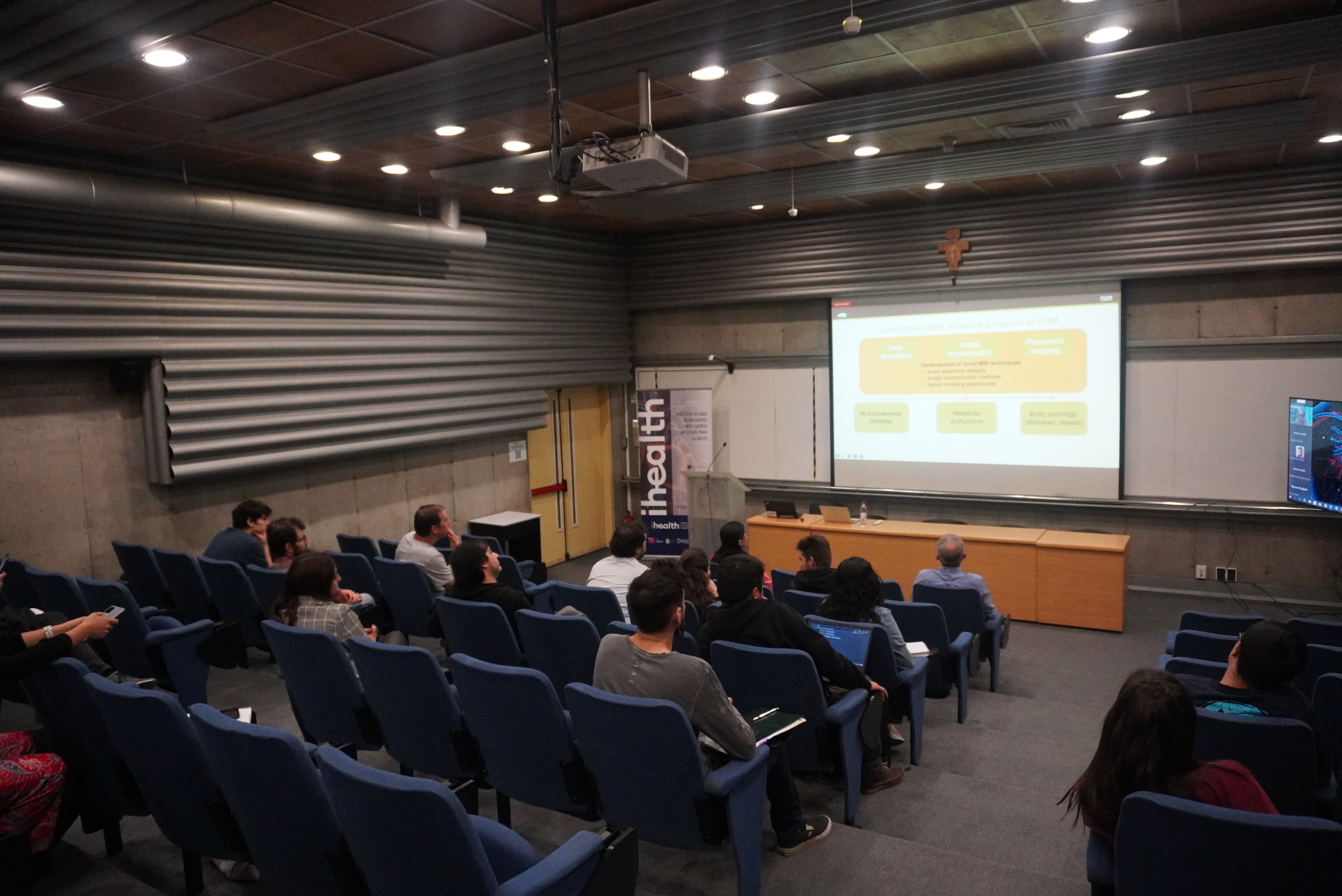
BIO: Prof. Karampinos’ research focuses on the development of new methods for magnetic resonance imaging (MRI). His research group develops MRI acquisition, image reconstruction and signal modeling techniques in order to generate new quantitative MRI biomarkers and to increase the robustness and effectiveness of quantitative MRI biomarkers. The developed methods are being translated into clinical studies for improving the diagnosis, the therapy monitoring, and the understanding of disease pathophysiology in musculoskeletal disorders, in metabolic diseases and in body oncology.
Prof. Karampinos studied engineering at the National Technical University of Athens, Greece. In 2008, he obtained his PhD with a focus on biomedical engineering from the University of Illinois, Urbana-Champaign. Between 2009 and 2012, he was a Postdoctoral Scholar in the Department of Radiology and Biomedical Imaging in University of California, San Francisco. He joined the Department of Diagnostic and Interventional Radiology of TUM in 2012 as a Junior Group Leader and he was appointed to the TUM School of Medicine in 2019.
Cristóbal Arrieta
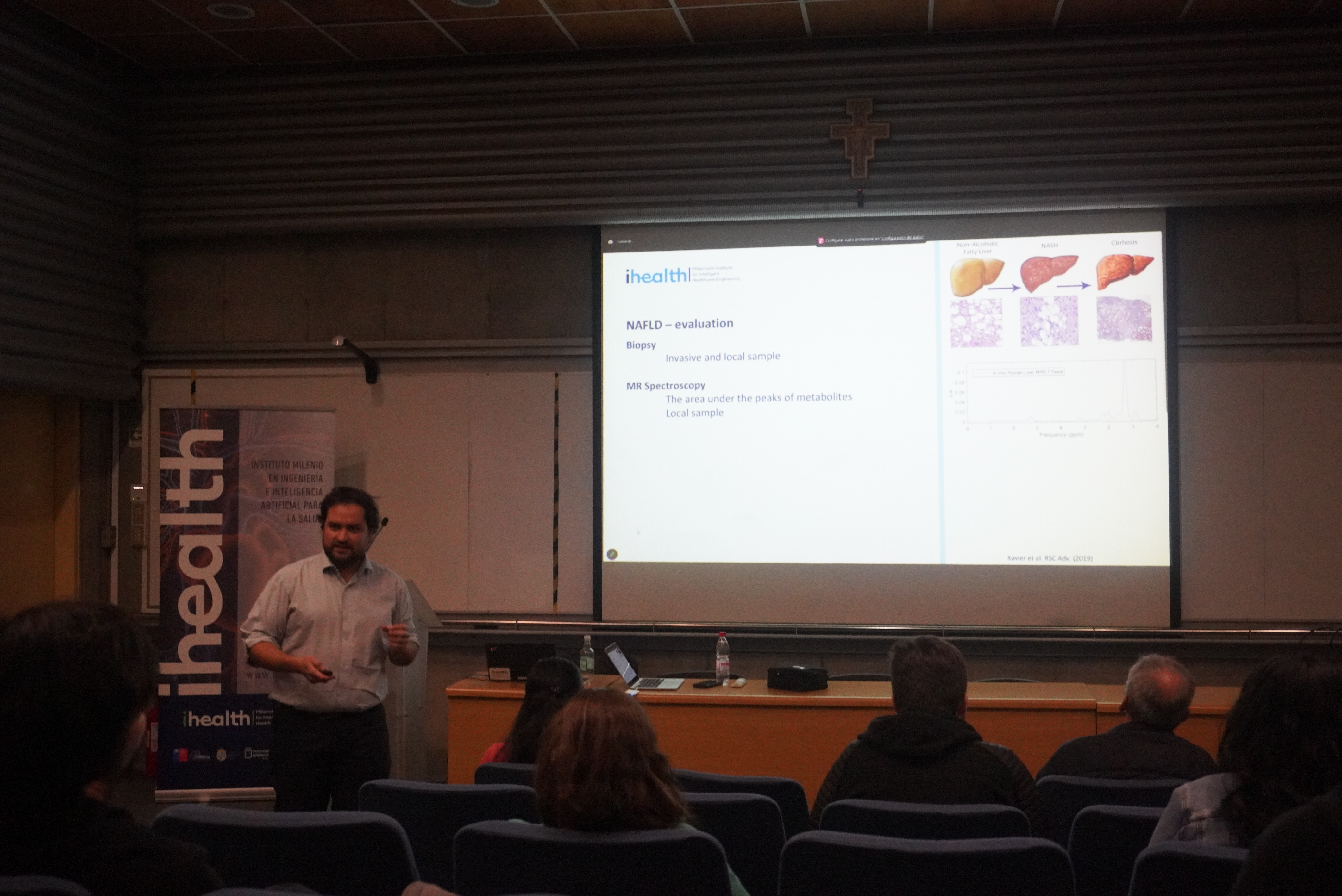
TITLE: “Magnetic Resonance Imaging Proton Density Fat Fraction (MRI-PDFF): accomplished promises and remaining challenges of this Biomarker for Non-alcoholic fatty liver disease”
ABSTRACT: Magnetic Resonance Imaging Proton Density Fat Fraction (MRI-PDFF) is a biomarker for quantifying fat. MRI-PDFF is especially useful for diagnosing Non-Alcoholic Fatty Liver Disease (NAFLD) and following up with NAFLD patients. MRI-PDFF was formally introduced in 2012, with a great effort of the scientific community to collaborate and share experiences with high levels of transparency to generate a consensus. The outcome of this consensus allows for fast and complete validation of MRI-PDFF across centers, scanner vendors, MRI sequences and parameters, and algorithms to fulfill the requirements for a standardized biomarker: It must be accurate, precise, robust, and reproducible.
This talk will discuss how the scientific community accounted for all these terms until the FDA approved commercially available solutions in 2017. We will review the main sequences, models, algorithms, and confounder factors to be corrected for computing a standardized MRI-PDFF. We will also discuss the remaining challenges and improvements, including some projects our team is working on.
BIO: Cristobal Arrieta is an academic of the new Faculty of Engineering at Universidad Alberto Hurtado. He got his Ph.D. at Pontificia Universidad Catolica, and his work was focused on medical image segmentation with prior knowledge. He is currently working on improving non-invasive liver fat quantification using MRI.